# 小鼠肠癌造模
## 动物实验基本原则
示例:All animal experiments were approved by the Institutional Animal Care and Research Advisory Committee of XXX in compliance with all relevant ethical regulations. Both male and female mice were used. Mice were housed under a regimen of 12 h light:12 h dark in non-specific pathogen-free (conventional) facility with a constant supply of food and water. Temperature was checked daily and maintained at 22 ± 2 °C, humidity was checked daily and maintained between 45–70%. Sample size was determined using power calculations with B = 0.8 and P < 0.05 based on preliminary data and in compliance with the 3R system: replacement, reduction, refinement. All mice were randomized before injections and samples were analysed in a blinded manner.(参考文献:Aspartate signalling drives lung metastasis via alternative translation)
肿瘤大小:Primary tumour volumes (calculated using the following formula: volume = (π/6) × length × width × height) were measured during every experiment using a manual calliper and weighted at the end of the experiment. During the course of the experiments, mice were monitored for detection of humane end points, determined using a score sheet (tumour size of 1.8 cm3, loss of ability to ambulate, laboured respiration, surgical infection or weight loss over 10% of the initial body weight). For all experiments the tumour volume did not exceeded 1.8 cm3.(部分文献伦理要求为1500mm3)(参考文献:Aspartate signalling drives lung metastasis via alternative translation)
实验终点:Regarding experimental endpoints, the maximum tumour size for subcutaneous tumours was a linear measurement of 2 cm at the largest diameter. For mice undergoing intrasplenic or spontaneous tumorigenesis induction, a loss of 20% of body weight, distress, hypothermia, bowel obstruction, severe hunched posture or inactivity were considered potential endpoints. Mice were humanely euthanized according to IACUC Euthanasia guidelines. Endpoint criteria were strictly followed in all animal experiments.(参考文献:Modeling resistance of colorectal peritoneal metastases to immune checkpoint blockade in humanized mice)
## 小鼠编号
### 耳标器编号
耳标器编号法常适用于成年小鼠,除特殊表型如先天性耳朵溃烂的品系不适用外其他品系均适用。操作人员单手或双手保定好小鼠,露出小鼠的耳朵,使小鼠腹部向下。将带有编号的耳标装入耳标器内,使用耳标器将耳标打入小鼠的耳朵上,将耳标有编号的一侧置于耳后,便于查看。
### 断趾编号
## 1、原位瘤模型
### 1)皮下荷瘤
参考文献:Modeling resistance of colorectal peritoneal metastases to immune checkpoint blockade in humanized mice
总体描述:For subcutaneous tumour cell inoculation, 8-week-old nude mice were anaesthetized with isoflurane. Cells were resuspended in growth factor-reduced Matrigel (Corning) and injected subcutaneously to the right flank using a 27 G needle. Tumour size was estimated with the formula: volume = width × width × length/2.
### 2)直肠原位荷瘤
### 3)PDO皮下荷瘤
* 参考文献:Modeling resistance of colorectal peritoneal metastases to immune checkpoint blockade in humanized mice
* 类器官使用消化酶消化为单细胞后,300G离心3min,培养基重悬计数(每侧肿瘤5\*10^5个细胞),300G离心3min,使用1:1的基质胶和培养基重悬,注射于NSG小鼠的皮下。
## 2、转移瘤模型
### 1)脾脏注射肝转移模型(Intrasplenic injection of tumor cells followed by splenectomy)
参考文献:Histone demethylase KDM5D upregulation drives sex differences in colon cancer
总体描述:For intrasplenic injection, 8-week-old nude mice were anaesthetized with isoflurane. The spleen was exteriorized through a left lateral flank incision. Cells resuspended in growth factor-reduced Matrigel (Corning) were injected using a 27 G needle. The injection site was pressed with a cotton stick for 1 min after withdrawing the needle to minimize cell leakage. The peritoneum was closed with suturing and skin was closed with wound clipping.
### 2)尾静脉/眶后静脉注射肺转移模型
参考文献:Zeb1 mediates EMT/plasticity-associated ferroptosis sensitivity in cancer cells by regulating lipogenic enzyme expression and phospholipid composition
总体描述:100 μl PBS containing 5 × 10⁴ cells were injected into the tail vein of 8-week-old C57BL/6NRj mice ( Janvier Labs). Littermates of both sexes were randomized for all treatment cohorts, monitored twice per week and killed 3 weeks after injection. Lungs were isolated, fixed in 4% paraformaldehyde and embedded in paraffin. Lung tissues were sectioned at 4-μM thickness and stained with haematoxylin and eosin solution. Per mice, metastatic lesions were screened on three sections separated by at least 200 μm. Quantification was performed by analysing the number of metastases as well as metastatic areas normalized to the respective lung area using ImageJ v.1.53a. For each treatment condition, nine mice were used in three independent experiments. The number and size of metastases never exceeded the maximal burden permitted by the local authorities.
1、术前准备
* 术前1天:动物房传入小鼠麻醉剂(三溴乙醇)、小鼠耳标、耳标钳、1毫升注射器、胰岛素针(0.25\*8mm,U-40(1毫升40小格,每小格25μL));
* 计算需要的肿瘤细胞量并铺板;
2、消化细胞
* 提前准备肿瘤细胞,术前胰酶消化,200G离心3min;
* 用8mL 预热的PBS重悬细胞,计数,再离心;
* 用PBS重悬至1\*10^6个/mL(细胞注射量需要预实验摸索,HCT116注射NSG按每只小鼠100μL(1\*10^5个)准备细胞,并多准备20%的余量)
3、术前准备
* 将细胞悬浮液放在冰上;
* 提前拿出并打开加热垫,酒精棉球擦拭;
* 对小鼠进行称重并记录;
* 根据小鼠体重,腹腔注射0.2 mL/10g 体重的三溴乙醇,麻醉小鼠(20g小鼠三溴乙醇用量在350-400μL之间);
* 用耳标进行标记,并记录分组;
4、手术操作
* 颠倒混匀细胞悬液,用胰岛素针吸取100μL细胞悬液;
* 固定小鼠头部,并使眼球突出于眼眶,进行眼眶后注射(Retro-orbital injections);
* 注射前针尖(尖口朝向内侧,以保护眼球不被划伤)与小鼠鼻尖方向呈30°角缓慢刺入内眦,深度为2\~3mm,见注射器有回血即可进行注射。注射时缓慢推注,结束后缓慢退针;
{% embed url="" %}
5、取材操作
* 注射3周后,处死小鼠,取肺组织,用4%PFA固定,制作石蜡切片;
* 每个蜡块切3刀,相隔200μm(50刀)以上,切成4μm薄片,进行HE染色并扫片;
* 对转移灶数量以及转移面积进行定量(相对于肺切片面积);
### 3)盲肠原位注射自发肝转移模型
{% file src="" %}
{% embed url="" %}
#### 1、术前准备:
* 术前2天:传入小鼠麻醉剂(三溴乙醇)、小鼠耳标、耳标钳、脱毛膏、1毫升注射器;
* 术前1天:
* 麻醉小鼠后,标记耳标以及腹部脱毛;
* 传入胰岛素针(0.25\*8mm,U-40(1毫升40小格,每小格25μL))、手术器械(直剪、弯剪、镊子、持针器若干)、手术缝线、AutoClip连续缝合器及缝合钉、棉签、小鼠加热垫;
#### 2、消化细胞
* 提前准备2个T75或10cm皿的肿瘤细胞,术前胰酶消化,200G离心3min;
* 用8mL 预热的PBS重悬细胞,计数,再离心;
* 用PBS重悬至4\*10^7个/mL(一个T75的HCT116长满约1\*10^7,用250μL PBS重悬)【按每只小鼠50μL(2\*10^6个)准备细胞,并多准备20%的余量】
3、术前准备
* 将细胞悬浮液放在冰上;
* 提前拿出并打开加热垫,酒精棉球擦拭;
* 对小鼠进行称重并记录;
* 根据小鼠体重,腹腔注射0.2 mL/10g 体重的三溴乙醇,麻醉小鼠(20g小鼠三溴乙醇用量在350-400μL之间);
* 用酒精或碘伏对小鼠腹部进行消毒;在手术部位周围放置无菌纱布,防止污染;
4、手术操作
* 使用镊子和手术剪刀,打开腹部外层及内部肌肉层;
* 使用镊子和棉签识别并暴露盲肠(盲肠通常位于胃的右侧),不要用镊子拖动组织;
* 颠倒混匀细胞悬液,用胰岛素针吸取50μL细胞悬液;
* 用棉签固定注射部位(避开血管),将胰岛素注射器的针头针尖朝上,轻轻插入浆膜下层(约1毫米);
* 将50μL细胞悬液缓慢注入半透明浆膜下层,并握住注射器一段时间以防止泄露。如果注射成功,将观察到浆膜下肿胀;
* 检查注射部位是否泄漏和出血,用生物胶水封闭肠壁防止泄露,将盲肠轻轻还纳入腹腔;
* 使用无菌缝合线和持针器,用不连续的结闭合内部肌肉层,用AutoClip缝合皮肤层;
5、术后处理
* 给小鼠注射50 μL的地塞米松(0.1 mg/mL),以避免手术引起的炎症,持续3天;
* 术后密切观察伤口愈合情况,记录体重;
* 4-6周后小鼠将死亡;
* 创建一个excel文件来记录每只鼠标的数据。要记录的数据应包括性别、出生日期、年龄和手术前的体重,以及终点的体重。在终点,应记录肿瘤重量、肝转移数量、原发性肿瘤和肝脏的照片。
### 4)盲肠原位植入PDO自发转移模型(murine orthotopic cecum-implantation model)
* 在植入前一天,将肿瘤类器官分离成单细胞,并将2.5×10^5个细胞接种在10μL大鼠尾高浓度I型胶原滴中
* 通过腹部中线切口切开盲肠,并将含有类器官的单个胶原蛋白滴手术移植到盲肠黏膜下层
{% hint style="info" %}
参考文献:Modeling resistance of colorectal peritoneal metastases to immune checkpoint blockade in humanized mice
A surgical orthotopic organoid transplantation approach in mice to visualize and study colorectal cancer progression
{% endhint %}
### 5)结肠镜引导下的类器官粘膜下注射结直肠癌转移模型(Colonoscopy-guided submucosal injection of CRC organoids)
* 传代后 36 小时,机械解离 AKPS 或 APTAK 类器官,重悬于 OptiMEM中(对于 AKPS,每只小鼠在 50 μl OptiMEM 中放入 1 个 50 μl Matrigel 圆顶;对于 APTAK,每只小鼠在 50 μl OptiMEM 中放入 3 个 50 μl Matrigel 圆顶) )。
* 麻醉小鼠并将其仰卧在加热垫(37°C)上。
* 使用安装在 50 ml 注射器上的血管内导管上的塑料管,用37 °C预热的 PBS 将结肠中的粪便排出。 使用定制注射针(33 gauge, 400 mm length, point style 4, 45° angle, Hamilton)、注射器,和带有集成工作通道的结肠镜(Storz)注射类器官溶液。
* 将针头接触结肠粘膜,快速输送50μl类器官溶液,形成粘膜下注射泡。 然后对小鼠进行监测,直到达到实验或人道终点。
{% hint style="info" %}
参考文献:
In vivo interaction screening reveals liver-derived constraints to metastasis;
Colonoscopy-based colorectal cancer modeling in mice with CRISPR–Cas9 genome editing and organoid transplantation
{% endhint %}
### 5)类器官脾脏注射转移模型
参考文献:
* In vivo interaction screening reveals liver-derived constraints to metastasis;
* Transplantation of engineered organoids enables rapid generation of metastatic mouse models of colorectal cancer
实验步骤:
* 前期准备:
* 将手术部位的皮肤剃毛并用碘伏/酒精消毒;
* 所有手术器械在使用前均经过消毒,手术过程在无菌条件下进行。
* 类器官消化:
* 在37°C下,在0.25%胰蛋白酶/0.02%EDTA中消化10分钟后,将类器官片段制备为单细胞,然后进行机械解离。
* 用DMEM(+P/S+10%FBS)重选,290g离心3 分钟,然后重新悬浮在100%PBS中,计数。
* 麻醉诱导后,在无菌条件下,对动物腹部进行剃毛和消毒。做一个1厘米的侧腹切口,露出脾脏尖端,将其从腹部轻轻抽出,并用无菌Q-tip固定到位。
* 脾脏注射:
* 使用无菌器械,在皮肤和腹膜壁上切开切口以暴露脾脏,将其从腹部轻轻抽出,用无菌枪头固定到位。 将无菌纱布放置在脾脏下方。
* 使用胰岛素注射器(0.3ml,30G),在30秒内将含有300000个细胞的0.1 mL总体积注射到脾包膜下。
* 拔出针头后,保持压力3-5分钟以确保无出血,然后切除脾脏,关闭切口。
* 用无菌PBS清洗伤口3次,并用缝线/伤口夹封闭皮肤。
* 监测小鼠的体重减轻,并在注射肿瘤细胞后最多3周终止实验。
## 3、自发成瘤模型
### 1)WT小鼠或肠上皮特异性敲除鼠(如XXXfl/fl-VillinCreERT2(xxxΔIEC)) + AOM/DSS诱导
原理:AOM/DSS模型是目前使用最广泛的UC化学诱导模型,临床症状学、形态学及病理学观察证明,该模型与人类溃疡性结肠炎癌变过程极为相似。AOM引起鸟嘌呤O6甲基化是诱导基因突变损伤最主要的原因,基因突变使细胞过度增殖,形成特异性的大肠肿瘤。DSS的致炎机制尚未阐明,可能与DSS的负电荷影响 DNA 合成、抑制上皮细胞增生、破坏肠黏膜屏障、导致巨噬细胞功能障碍及肠道菌群失调有关。
* 第1天称重并标记小鼠,腹腔注射AOM(10mg/kg体重(或12.5mg/kg);SigmaAldrich/MCE)
* 1周后进行三个周期的DSS给药。每个DSS处理周期包括7天含2.5%DSS的水,然后是14天的常规饮用水。
* 重复三个周期。
{% hint style="info" %}
AOM配置方法:首先将AOM溶于生理盐水,0.22μm过滤,配成10mg/mL贮存液(分装后保存于-20℃),使用之前,融化分装母液并用无菌生理盐水10倍稀释。配成1mg/mL。20g小鼠体重注射200μL。
DSS配置方法:先用无菌dd水配置10%DSS贮存液(比如10g溶于100mL)。用50mL离心管传进动物房后,用150mL ddH2O 1:3稀释至200mL。动物房申请红色纸,标注浓度日期,标明不要换水。隔天更换一次DSS溶液。
{% endhint %}
### 2)ApcMin/+小鼠 (germline Apc mutation,主要为小肠腺瘤)
* 自发成瘤
### 3)Apcfl/fl×VillinCreERT2小鼠 + 4-OHT(Apc-null adenomas)
* 4周龄时,用4-羟基他莫昔芬(4-OHT,1mg/mL;Sigma-Aldrich)灌肠,将1cm的直肠管插入肛门,并给予50μL 4-OHT溶液
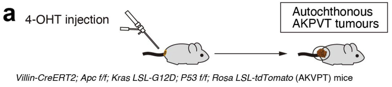
### 4)iAKP小鼠+ 4-OHT灌肠(Autochthonous tumour models)
* 肠上皮条件性特异性缺失APC、Trp53及KRAS突变:VillinCreERT2;Apcfl/fl;KrasLSL-G12D;Trp53fl/fl
* 4周龄时,用4-羟基他莫昔芬(4-OHT,1mg/mL溶于无水乙醇;Sigma-Aldrich)灌肠,将1cm的直肠管插入肛门,并给予50μL 4-OHT溶液,6周后开始出现原位结直肠癌
{% hint style="info" %}
4-OHT操作
一、4‑OHT给药方案(频率、次数、体积/剂量)
\
常规方案:每日1次,连续3天(q.d. × 3)。
\
剂量与体积(按体重与浓度双控)
\
剂量:1–2 mg/kg/次(多数情况下1.5 mg/kg 已足够诱导肠上皮CreER)。
\
体积:100–150 μL/只(20–25 g小鼠)。避免>200 μL以减少逆流与腹压不适。
\
工作液浓度:5–10 mg/mL(更高浓度可使体积更小、减少逆流,但溶剂比例也更“刺激”,需平衡)。
\
灌药前4–6小时禁食但不禁水,有助于减少粪便干扰与逆流。
\
给药后将小鼠头低臀高(轻微Trendelenburg位)保持3–5分钟,帮助药液留置。
\
二、4‑OHT溶解与载体
4‑OHT粉末先溶于无水乙醇或DMSO,配制20–50 mg/mL的储备液,-20°C避光保存(反复冻融≤3次)。
\
灌肠用工作液(不含水)
\
常用载体:玉米油(corn oil)或葵花籽油。操作是“储备液(EtOH或DMSO)慢慢滴加入油相中,涡旋充分、37°C水浴震荡10–15分钟”形成均一悬/溶液。
\
最终有机溶剂比例控制:EtOH或DMSO不超过2%(v/v),总有机相低可减少黏膜刺激。
\
三、灌肠操作细节(关键影响复现度)
\
插入深度:3–4 cm(成年小鼠)。过浅易逆流,过深有穿孔风险。操作时顺直肛门方向轻柔推进,切勿用力。
\
导管尖端涂少量无菌凡士林或水溶性润滑剂;工作液避免产生气泡,空气一并注入会诱发排便反射。
\
缓慢推注(10–20 μL/秒),推注完毕停留5–10秒再撤管。
\
撤管后让小鼠保持头低臀高或竖握姿势3–5分钟,观察是否有回流。
三、其他
麻醉与应激
\
一般不需全麻,轻度异氟烷吸入可降低抵抗、减少损伤;操作时间尽量<1分钟。
\
术后护理
\
放回干燥洁净垫料,单笼或小群饲,1–2小时内观察是否出血、显著不适或持续腹泻。
\
感染/炎症控制
\
全程无菌操作,导管一次性或严格消毒;连续多天给药时,必要时间隔给益生元/等渗电解质以降低黏膜刺激(依情况而定)。
{% endhint %}
参考文献:Oncogenic Kras drives invasion and maintains metastases in colorectal cancer
* 如果AKP小鼠进一步杂交Rosa26LSL-tdTomato ,可以得到自发生成荧光标记肿瘤的AKPVT小鼠
* 如有条件,可采用内镜下注射50μL 100μM 4-OHT(溶于PBS)的操作方式
SOX17 enables immune evasion of early colorectal adenomas and cancers
* AKP小鼠进一步杂交Tgfbr2fl/fl得到AKTP小鼠
## **他莫昔芬给药**
* 配置方法:在50mL离心管中称量**Tamoxifen**粉末(Sigma‐Aldrich,# 10540-29-1),溶于玉米油(Sigma‐Aldrich,# 8001-30-7)至20mg/mL,摇床震荡过夜溶解;
* 全身给药方法:腹腔注射给药,100mg/kg(小鼠体重20g,约给予100μL/只),连续给5天。
{% file src="" %}
## 多西环素给药
* 饲料:doxycycline food pellets (2,500 mg kg–1; Envigo)
* 饮水:doxycycline (200 μg/mL加至饮水,每周更换)
## 生物发光成像(BLI)
* **工作液配置:**
* 溶解 1 g荧光素钾盐或钠盐于 66.6 ml 无Ca2+和Mg2+的DPBS中,配置成 **15 mg/mL**的溶液。
* 混匀,0.2μm滤膜过滤(可吹入惰性气体,如氮气,以防止氧化。)-80℃避光保存,一年内有效。
* **使用:**
* 在冰上避光解冻(若是在冰上比较难解冻,可短暂温浴,并轻柔混匀)。
* 按照动物体重 **150 mg/kg**进行注射,(例如,20 g重的小鼠需要注射腹腔注射200μl )
* 注射入体内10-20 min(待光信号达到最强稳定平台期),再进行成像分析。
注:建议对每只动物模型都需要建立荧光素酶动力学曲线,从而确定最高信号检测时间和信号平台期。
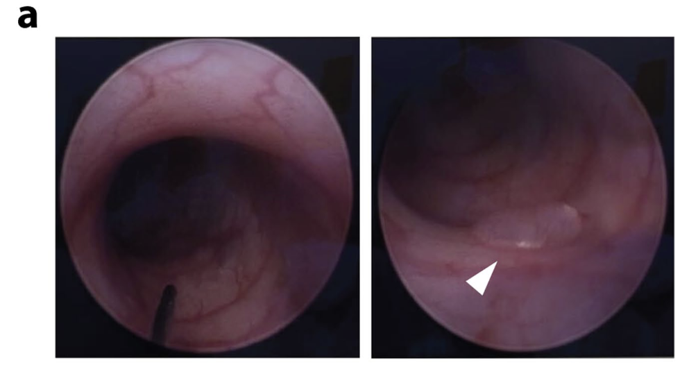
## 小鼠肠镜
* For spontaneous CRC mouse models, mouse colonoscopy was performed with KARL STORZ ENDOSKOPE (length: 10cm, diameter: 19mm). Mice were fasted at least 8h on the day prior to colonoscopy. After anaesthesia by inhalation of 4% isoflurane via Gas Filter Canister, the endoscope was introduced from the mouse anus with CO2 insufflation (pressure less than 10-15 mmHg, gas flow 5-10L/min) to keep the colon inflated. Colon was rinsed with warm PBS during pushing the endoscopy to the point of colon curve if necessary. Endoscopy will be slowly withdrawn and started to capture images until reaching anus. All procedures were approved by the Animal Experimentation Ethics Committee.
* 参考文献:Peptostreptococcus stomatis promotes colonic tumorigenesis and receptor tyrosine kinase inhibitor resistance by activating ERBB2-MAPK
## 小鼠自发肠癌的病理评分
### 1、Histology assessment
* Intestinal lesions in murine models are characterized by abnormal/dysplastic epithelial maturation with an increasing nucleus–cytoplasm ratio, hyperchromatic, enlarged, and irregular nuclei populating the crypts, which can be further divided into low-, high-grade dysplasia and adenocarcinoma.
* In low-grade dysplasia, crypts are crowded and arranged in parallel with crowded penicillate nuclei retained at the basal aspect.
* High-grade dysplasia is characterized by architectural complexity including cribriform glandular structures, back-to-back or solid nest-like structures along with luminal necrosis, while cytologic atypia is prominent with large nuclei, loss of polarity and numerous mitoses, but without definite stromal invasion.
* Adenocarcinoma is characterized by irregular glands with nuclear pleomorphism and loss of nuclear polarity, lamina propria/muscularis mucosae invasion and infiltration
### 2、Inflammation scoring
* Score 0, no inflammation;
* Score 1, mild inflammatory with occasionally present occasional inflammatory cells;
* Score 2, moderate inflammatory;
* Score 3, severe inflammation;
* Score 4, critical inflammation with multiple crypt abscesses
{% hint style="info" %}
参考文献:
* Peptostreptococcus stomatis promotes colonic tumorigenesis and receptor tyrosine kinase inhibitor resistance by activating ERBB2-MAPK
* 病理学分级:Characterization of Colorectal Cancer Development in Apc (min/+) Mice;Pathology of mouse models of intestinal cancer: consensus report and recommendations
* 炎症评分:A guide to histomorphological evaluation of intestinal inflammation in mouse models
{% endhint %}